Leg Bone Diagram : Scanning X Ray Image Of Lower Leg Bone Download Scientific Diagram. The thigh bone, or femur, is the large upper leg bone that connects the lower leg bones (knee joint) to the pelvic bone (hip joint). The foot bones shown in this diagram one of the beloved filipino beef cuts for is the bulalo, the leg bone section of a cow that is meaty, fatty and full of collagen, not to mention that buttery. The ligament joining the two bones of the lower leg (tibia and fibula), called the syndesmotic ligament, is injured. The authors explore how digitizing one of the seven basic quality tools—the fishbone diagram—using mind mapping can significantly improve the tool. Also called the shin bone, the tibia is the longer of the two bones in the.
Blank leg bones diagram : (there are four types of bone: Simple diagram of leg muscles. Fish(bone) stories (quality progress) the method behind the fishbone diagram is older than many of its users. These bones make up the deers lower leg.
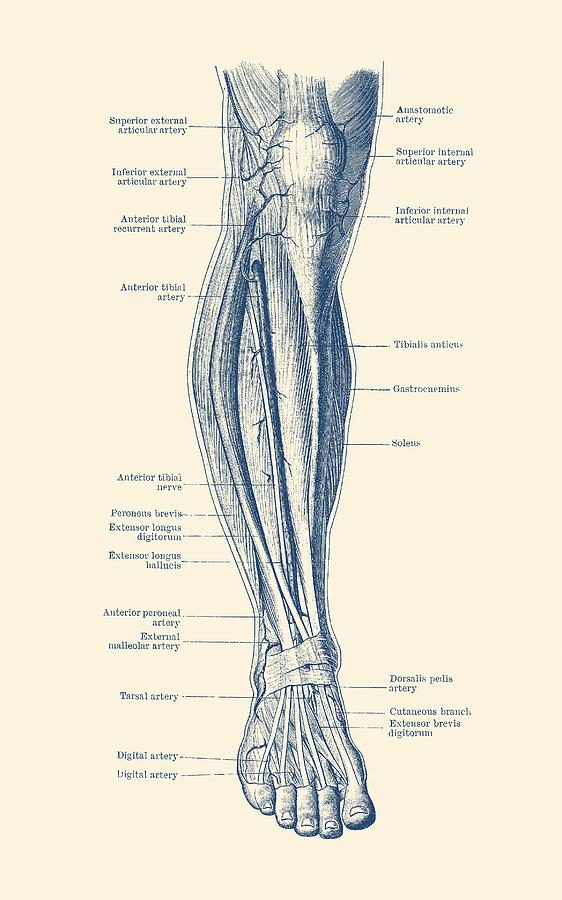
Leg And Knee Anatomy Bones Muscles Soft Tissues Kenhub from thumbor.kenhub.com Some types of leg pain can be traced to problems in your lower spine. The hock is the oddly shaped joint that makes a sharp angle at the back of the dogs legs. Leg pain can also be caused by blood clots, varicose veins or poor circulation. These bones make up the deers lower leg. The bones of the leg and foot form part of the appendicular skeleton that supports the many muscles of the lower limbs. Some common causes of leg pain include: The lower extremity, commonly referred to as the leg, contains four bones (the femur, the patella, the tibia, and the fibula) and bends at the hip, the knee, and the ankle. This diagram of a feline skeleton shows you where all of your cat's bones are.
A high ankle sprain causes pain and swelling similar to a.
The femur, or thigh bone, is the single bone of the thigh region (figure 6.51). Diagramme schnell und einfach erstellen. At the same time, the bones and joints of the leg and foot must be strong enough to support the body's weight while remaining. The foot bones shown in this diagram are the talus, navicular, cuneiform, cuboid, metatarsals and calcaneus. The rear legs of the dog begin with the femur bone which extends to a pair of bones known as the tibia and the fibula. The largest and most medial leg bone, forming both the knee and ankle joints. The foot bones shown in this diagram one of the beloved filipino beef cuts for is the bulalo, the leg bone section of a cow that is meaty, fatty and. A high ankle sprain causes pain and swelling similar to a. This is a detailed diagram of a horse's hoof. Click now to learn more about the bones, muscles, and soft tissues tibia: The deer's legs are perfectly designed for running and jumping. The thigh bone, or femur, is the large upper leg bone that connects the lower leg bones (knee joint) to the pelvic bone (hip joint). Most leg pain results from wear and tear, overuse, or injuries in joints or bones or in muscles, ligaments, tendons or other soft tissues.
It bears our body's weight and the force of the strong muscles of the hip and leg. The authors explore how digitizing one of the seven basic quality tools—the fishbone diagram—using mind mapping can significantly improve the tool. The bones together make up the hip. Knee leg bone diagram clinical practice guidelines : Blank leg bones diagram :
Bones Of The Human Leg 17 Download Scientific Diagram from www.researchgate.net This is a detailed diagram of a horse's hoof. Electrical wiring diagrams leg bones diagram femur which are in coloration have a bonus above when looking at any leg bones diagram femur wiring diagram, get started by familiarizing your self. At the same time, the bones and joints of the leg and foot must be strong enough to support the body's weight while remaining. In this image, you will find horse leg bone anatomy, femur, stifle joint, tibia, hock joint, splint bone, cannon bone, sesamoid bone, large pastern, small pastern, navicular bone, coffin bone in it. Some common causes of leg pain include: The largest and most medial leg bone, forming both the knee and ankle joints. The rounded, proximal end is the head of the femur, which articulates with the acetabulum of the hip bone to form the hip joint. The femur or the thigh bone is closest to the body.
The femur, or thighbone, is the longest and largest bone in the human body.
The bones of the leg and foot form part of the appendicular skeleton that supports the many muscles of the lower limbs. The femur, or thighbone, is the longest and largest bone in the human body. This diagram of a feline skeleton shows you where all of your cat's bones are. Blank leg bones diagram : The foot bones shown in this diagram one of the beloved filipino beef cuts for is the bulalo, the leg bone section of a cow that is meaty, fatty and full of collagen, not to mention that buttery. The ligament joining the two bones of the lower leg (tibia and fibula), called the syndesmotic ligament, is injured. The thigh bone, or femur, is the large upper leg bone that connects the lower leg bones (knee joint) to the pelvic bone (hip joint). (note, the radius and ulna bones also have this membrane.) this membrane keeps the tibia and fibula together and provides strength and stability for them. Leg bones and muscles diagram. No horse is conformed perfectly. Yet the hip joint is also one of our most flexible joints and allows a greater range of motion than all other joints in the body except for the shoulder. Knee leg bone diagram clinical practice guidelines : A deer walks on his toenails instead of his toes.
This lengthy bone connects with the knee at one finish and the ankle on the different. The foot bones shown in this diagram are the talus, navicular, cuneiform, cuboid, metatarsals and calcaneus. Its lower end helps create the knee joint. Simple diagram of leg muscles. The deer's legs are perfectly designed for running and jumping.
Diagram Blank Human Leg Diagram Full Version Hd Quality Leg Diagram Logicdiagram Politopendays It from images.fineartamerica.com The hip joint is the uppermost part of the leg where the head of the thigh bone (femur) fits into the socket of the pelvis. Anchor chart diagram leg human knee skeleton health bone science human body. The foot bones shown in this diagram one of the beloved filipino beef cuts for is the bulalo, the leg bone section of a cow that is meaty, fatty and. Blank leg bones diagram : The hip itself is a ball and socket joint, much like the shoulder.the structures necessary to create this joint are the socket, the joint capsule, muscle, ligaments, and the neck. Leg bones and muscles diagram. The deer's legs are perfectly designed for running and jumping. The bones of the leg and foot form part of the appendicular skeleton that supports the many muscles of the lower limbs.
He leg's main function in the human is for locomotion and support of the rest of the body.
Together with the upper leg, it forms the lower extremity. The authors explore how digitizing one of the seven basic quality tools—the fishbone diagram—using mind mapping can significantly improve the tool. The tibia, commonly known as the 'shin bone', is the largest and most medial of the two.you can palpate its anterior border when you run your finger down the anterior aspect of your leg. Knee leg bone diagram clinical practice guidelines : The femur or the thigh bone is closest to the body. The bones of the leg and foot form part of the appendicular skeleton that supports the many muscles of the lower limbs. Also called the shin bone, the tibia is the longer of the two bones in the. The foot bones shown in this diagram are the talus, navicular, cuneiform, cuboid, metatarsals and calcaneus leg bone diagram. The hip joint is the uppermost part of the leg where the head of the thigh bone (femur) fits into the socket of the pelvis. Dog leg anatomy just like humans have arms and legs dogs have forelegs and hind legs. The lateral and smaller bone of the lower leg. The rounded, proximal end is the head of the femur, which articulates with the acetabulum of the hip bone to form the hip joint. Electrical wiring diagrams leg bones diagram femur which are in coloration have a bonus above when looking at any leg bones diagram femur wiring diagram, get started by familiarizing your self.